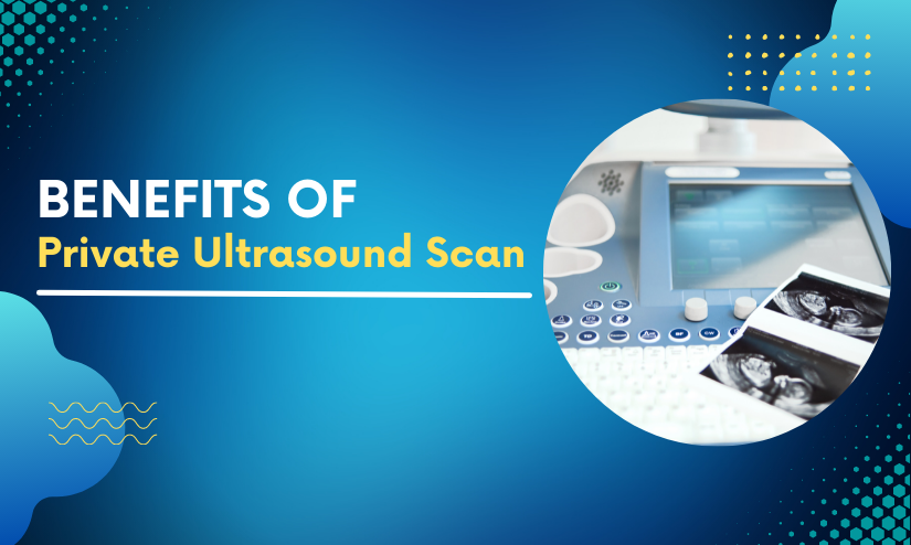
A Private Ultrasound Scan in Leicester is a simple technique that is often performed during pregnancy to get pictures of the growing baby. Follow our guide to discover all there is to know about prenatal ultrasounds and what they can do for you and your baby!
Are Obstetrical Ultrasounds Risky?
Ultrasound imaging is non-invasive and does not involve the use of radiation. For almost six decades, it has been used to assess pregnancy, and there has been no indication of damage to the patient, embryo, or baby. It should, however, only be done when medically required.
Ultrasound imaging offers a good image of the uterus as well as a wealth of information about your pregnancy. It may be unpleasant at first, but it should not be painful. While they might provide comfort regarding your baby’s health, we recognize that they can also generate concern.
Standard Ultrasound
An ultrasound, which is often used to confirm pregnancy during the first trimester, provides you with your first view of your developing baby. This first ultrasound will determine the health of your early pregnancy as well as confirm your due date.
Another ultrasound is generally conducted during the second trimester to picture all areas of your baby in depth; this is the period when the baby’s gender may be determined. This is a thrilling time for many parents, and they look forward to it with bated breath. Later in pregnancy, ultrasounds are used to assess fetal development and well-being.
Ultrasounds in 3D and 4D
3D and 4D scanning are useful approaches for detecting and monitoring possible issues during pregnancy. During a 3D ultrasound, you will be able to view a three-dimensional picture of your baby. The 4D ultrasound, like the 3D ultrasound, delivers a three-dimensional view of the baby as you see the images live.
The FDA advises that these ultrasounds be performed only by experienced certified diagnostic medical sonographers at their medical ultrasounds. Many sonographers will be able to supply you with this unique perspective as well as a memento photograph.
Fetal Echocardiography
If your doctor believes your kid has a congenital heart problem or if this medical condition runs in your family, this treatment will be performed. The echocardiogram provides a thorough picture of the baby’s growing heart, including its rhythm and blood flow. A fetal echocardiogram is typically conducted about 18 weeks of gestation; however, it may be performed as early as 12 weeks.